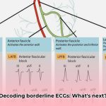
Ever wonder if those slightly unusual heart rhythm readings in young athletes are cause for concern? It’s a complex issue! A young athlete’s heart can look different on an ECG than a non-athlete’s due to the physiological adaptations resulting from intense training. This guide provides insights to help differentiate between normal variations and findings that warrant further attention. We’ll walk you through interpreting borderline ECGs – those readings that aren’t perfectly normal but aren’t obviously alarming. You’ll learn about the latest guidelines, practical tips for accurate interpretation, and discover best practices, enabling you to confidently care for your young athletes. Accurate assessment of ECGs is crucial for ensuring athlete safety and well-being. For more information on next steps, see this helpful guide on [what to do next](https://www.mearnes.com/what-to-do-after-a-borderline-ecg).
Borderline ECGs in Young Athletes: Interpretation and Risk Assessment
Decoding an electrocardiogram (ECG) for a young athlete requires careful consideration, especially when assessing potential cardiac risks. Borderline ECGs, results that fall into a “gray area”—neither clearly normal nor definitively showing a problem—demand meticulous interpretation. Misinterpreting these ECGs can have serious consequences, including unnecessary anxiety and disqualification from sports or, conversely, missing a potentially life-threatening condition. How can clinicians accurately assess these findings and ensure young athletes’ safety?
Athletic Hearts: Understanding Physiological Adaptations and ECG Changes
Regular, intense training induces structural and electrical remodeling of the heart, resulting in physiological adaptations. These adaptations often manifest as ECG changes, many of which are normal and reflect the heart’s response to the demands of exercise. For example, sinus bradycardia (a slower heart rate), sinus arrhythmia (slightly irregular beats), first-degree atrioventricular block (a prolonged PR interval), or incomplete right bundle branch block are common and often healthy in well-trained athletes. The challenge lies in distinguishing these normal adaptations from ECG patterns that may indicate underlying pathology. What specific ECG patterns should clinicians recognize as typical adaptations in athletes, and what are the underlying mechanisms driving these changes?
The Tricky Middle Ground: Spotting Possible Problems and Conduction Abnormalities
Differentiating between a “normal” athlete’s ECG and one that signals potential trouble requires expertise and specialized knowledge. A finding that appears abnormal in a non-athlete may be perfectly normal in a trained athlete. For example, early repolarization, characterized by ST-segment elevation, is frequently observed in athletes and is usually benign. However, recent studies have suggested a possible association between specific early repolarization patterns (e.g., J-point elevation in the inferior leads) and an increased risk of ventricular fibrillation in the general population. While the clinical significance of early repolarization in athletes remains controversial, careful evaluation is necessary to avoid both overdiagnosis and missed diagnoses. Do clinicians have clear guidelines for differentiating benign early repolarization from potentially dangerous patterns in athletes, considering factors such as age, sex, ethnicity, and sport?
The Whole Picture: Comprehensive Athlete Assessment Beyond ECG Interpretation
Interpreting a borderline ECG in a young athlete requires a holistic approach that extends beyond the ECG tracing. A comprehensive assessment includes a detailed medical history, assessment of training regimen, and physical examination. Key aspects of the medical history include:
- Family history of sudden cardiac death, inherited cardiac conditions, or arrhythmias
- History of syncope (fainting), near-syncope, or exertional chest pain or dyspnea
- Use of medications or illicit substances that may affect cardiac function.
The physical examination should focus on detecting signs of underlying cardiac disease, such as murmurs, abnormal heart sounds, or signs of heart failure. Additional tests, such as echocardiography (to assess cardiac structure and function) or exercise stress testing (to evaluate the heart’s response to exertion), may be necessary to further clarify the diagnosis and risk stratification. What specific historical and physical exam findings are most critical when evaluating a young athlete with a borderline ECG, and how should clinicians integrate these findings with the ECG data to guide further management?
Ethnicity and Age Considerations: Impact on ECG Patterns and Interpretation
Ethnicity and age significantly influence ECG patterns and interpretation in athletes. For example, Black athletes are more likely to exhibit certain ECG variations, such as T-wave inversions in the anterior precordial leads (V1-V4), which may be normal variants in this population. Similarly, the “juvenile T-wave pattern,” characterized by T-wave inversions in the right precordial leads (V1-V3), is a normal finding in adolescent athletes but may be abnormal in older individuals. Clinicians must be aware of these age- and ethnicity-related differences to avoid misinterpreting normal variations as pathological findings. How do age-related physiological changes and ethnic differences affect normal ECG intervals and morphology in young athletes, and what strategies can clinicians use to minimize the risk of misdiagnosis in these populations?
Practical Steps for Doctors and Trainers: Guidelines for Accurate ECG Assessment
Here’s a step-by-step approach clinicians can take:
- Start with the Basics: Always begin with a complete medical history and a thorough physical exam; this forms the foundation for everything else.
- Context is Critical: Remember that the athlete’s training regimen, ethnicity, and age all play key roles in interpreting the ECG; don’t interpret the ECG in isolation.
- Apply Standardized Criteria: Utilize established ECG interpretation criteria, such as the International Recommendations for ECG Interpretation in Athletes, to ensure consistent and accurate assessment.
- Consider Serial ECGs: When uncertainty exists, consider obtaining serial ECGs to assess for dynamic changes over time.
- When in Doubt, Test More: Don’t hesitate to order additional tests like echocardiograms or stress tests if the ECG results are unclear; it’s always better to be safe than sorry.
- Stay Up-to-Date: Keep abreast of the latest guidelines and research on ECG interpretation in athletes because medical knowledge constantly evolves.
- Teamwork Makes the Dream Work: If you’re unsure about an ECG interpretation, seek consultation from a cardiologist; obtaining a second opinion can provide reassurance or identify subtle abnormalities.
Looking Ahead: The Future of Borderline ECG Interpretation and Genetic Risk Factors
Our understanding of borderline ECGs in young athletes has improved, but further research is needed to refine diagnostic criteria and improve risk stratification. Future studies should focus on:
- Identifying genetic markers associated with increased risk of sudden cardiac death in athletes with borderline ECG findings.
- Developing more sophisticated algorithms for ECG interpretation that incorporate clinical, demographic, and genetic data.
- Evaluating the long-term outcomes of athletes with borderline ECGs to determine the optimal management strategies.
The ultimate goal is to screen athletes effectively without causing undue anxiety or unnecessary interventions. We need to strike a balance between caution and recognizing normal variations in athletic hearts. Will advances in genetic testing improve risk stratification for young athletes with borderline ECG findings, and how will these technologies be integrated into clinical practice?
Summary Table: Key Points to Remember for ECG Interpretation
| Feature | Important Considerations |
|---|---|
| Training Level | How hard and how long they train greatly influences the ECG. |
| Ethnicity | ECG patterns vary across different ethnic groups. |
| Age | What’s considered normal changes as athletes get older. |
| Family History | A family history of heart problems increases the risk. |
| Clinical Picture | Don’t just focus on the ECG; symptoms and other physical findings are vital. |
| ECG Criteria | Apply standardized criteria, such as the International Recommendations, for ECG interpretation. |
| Serial ECGs | Consider serial ECGs to assess for dynamic changes over time. |
Remember, responsible care means carefully investigating potential issues while also understanding the normal variations seen in the hearts of young athletes. It’s a delicate balance, but crucial for young athletes’ well-being. Is further training required to help healthcare professionals interpret borderline ECG findings in athletes, and what novel educational strategies can be implemented to improve ECG interpretation skills among clinicians?
How to Interpret Borderline ECG Findings in Black Athletes: A Clinical Guide
Key Takeaways:
- Normal ECG variations are common in athletes due to physiological adaptations to training; up to 25% of athletes may have ECG findings that require careful evaluation.
- How to interpret borderline ECG findings in Black athletes requires awareness of ethnic variations in ECG patterns, ensuring accurate diagnosis and avoiding overdiagnosis.
- A comprehensive assessment, including the athlete’s symptoms, family history, and other ECG features, is crucial for accurate interpretation and minimizing false positives.
- Further investigation may be necessary depending on the presence of additional abnormalities and the athlete’s clinical status, with echocardiography and exercise stress testing being valuable tools.
- Utilizing updated ECG interpretation guidelines and risk stratification tools is essential for appropriate management and improving athlete safety.
Understanding the Athlete’s Heart: Normal vs. Abnormal ECG Patterns
Interpreting electrocardiograms (ECGs) in athletes can be challenging. Athletic training causes significant changes to the heart’s structure and function, leading to ECG patterns that might seem concerning to the untrained eye. However, many ECG alterations are physiological adaptations, normal responses to intense physical conditioning. Conversely, certain findings raise red flags and demand thorough evaluation to rule out underlying cardiac conditions. Which specific ECG findings should prompt further comprehensive clinical and cardiac evaluation in athletes?
Recognizing Normal Adaptations and Physiological ECG Variations
Several common ECG findings represent normal physiological changes in athletes. These include:
*
- Best Mindfulness Books for Anxiety, Sleep, and Daily Peace - January 29, 2026
- Books On Mindfulness For A Happier, More Present Life - January 28, 2026
- Essential Meditation Books for Beginners and Experienced Practitioners - January 27, 2026


